HOW CAN WE HELP YOU? Call 1-800-TRY-CHOP
Single Cell Technology Core Tool & Fees
Technologies
10x Genomics Chromium Controller and Chromium X provides single cell whole transcriptome gene expression and multiplexing capabilities to profile hundreds to a million cells. Partitioning events occur on a microfluidic chip in the presence of barcoded gel beads and oil to create GEMs (Gel Bead in EMulsion) encapsulating single cells or nuclei. Illumina compatible libraries are constructed in bulk after GEMs are broken.
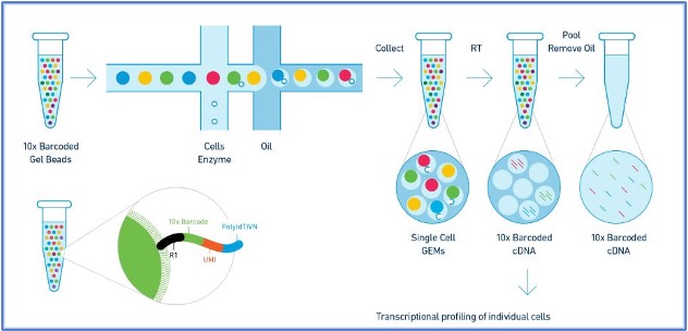
Source: 10xGenomic
For more information, please visit https://www.10xgenomics.com/technology
NanoString's GeoMx Digital Spatial Profiler (DSP) combines standard immunofluorescence techniques with digital optical barcoding technology to perform highly multiplexed, spatially resolved profiling of RNA and protein targets on tissue sections. Whole tissue sections, FFPE or fresh frozen, can be imaged and stained for RNA or protein. Specific tissue compartments or cell types can be selected to profile based on the biology, and the expression levels can be counted using either the nCounter Analysis System or an Illumina Sequencer.
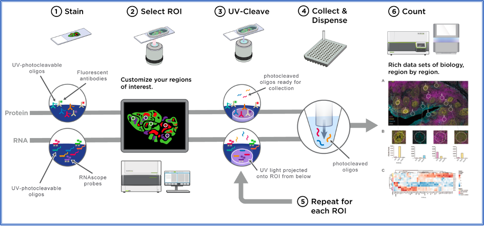
Source: nanoString
For more information, please visit https://nanostring.com/products/geomx-digital-spatial-profiler/geomx-dsp-overview/.
We offer highly multiplexed imaging using Akoya's PhenoCycler (formerly CODEX) platform. Through repetitive imaging cycles, this technology allows for the detection of a maximum of 40+ protein targets in the same tissue section at single cell resolution, with quantitative data for each protein.
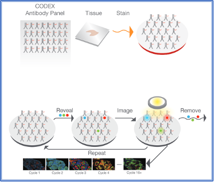
Source: Akoya Biosciences
For more information, please visit https://www.akoyabio.com/phenocycler/
Using combinatorial labeling, sequential imaging, and error-robust barcoding, MERFISH technology allows simultaneously measuring the copy number and spatial distribution of the transcripts of up to 500 genes in individual cells across whole tissue slices with subcellular resolution.

Source: Vizgen.com
For more information, please visit https://vizgen.com/resources/how-merfish-technology-works/
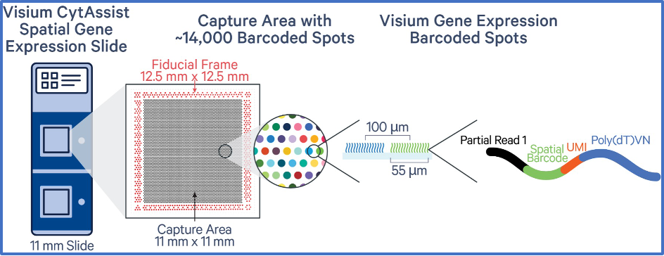
10x Genomics Visium CytAssist Spatial Gene Expression is a probe-based molecular profiling method that enables scientists to map whole transcriptome gene activity across an entire tissue section alongside H&E morphological imaging. The CytAssist facilitates the transfer of probes from tissues on standard glass slides to Visium slides—enabling truly unbiased spatial discovery across an expanding range of compatible tissues. Each Visium CytAssist Spatial Gene Expression Slide contains Capture Areas with barcoded spots that include oligonucleotides required to capture gene expression probes.
Source: 10xGenomic
For more information, please visit https://www.10xgenomics.com/spatial-transcriptomics
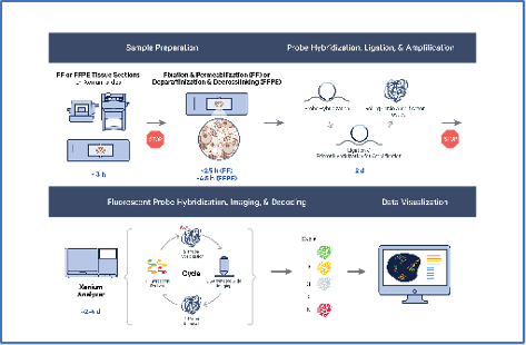
10x Genomics Xenium in situ platform uses a padlock probe rolling circle amplification chemistry. The tissue section is processed and selected probes hybridized to the RNA targets within the tissue. Bound probes are ligated, forming a circular template that allows rolling circle amplification of the probe and a unique barcode. Slides are placed in the Xenium Analyzer where the sample undergoes successive rounds of fluorescent labeling and imaging, creating a spatial map of the transcripts across the entire tissue section. Data is immediately viewable and is compatible with an easy-to-use data viewer and with many third-party downstream analysis tools, which continue to develop rapidly.
Source: 10xGenomic
For more information, please visit https://www.10xgenomics.com/in-situ-technology
