HOW CAN WE HELP YOU? Call 1-800-TRY-CHOP

In This Section
'New Technologies Lead to New Discoveries': Q&A With HHMI Scholar Mustafa Mir, PhD
Published on May 9, 2023 in Cornerstone Blog · Last updated 9 months ago
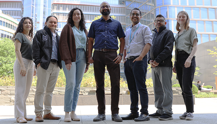
Mustafa Mir, PhD, shown here with his team, was selected as a Howard Hughes Medical Institute Freeman Hrabowski Scholar.
Editor's Note: Mustafa Mir, PhD, assistant professor in the Center for Computational and Genomic Medicine at Children's Hospital of Philadelphia, was selected as a Howard Hughes Medical Institute (HHMI) Freeman Hrabowski Scholar. This new program supports early career basic science faculty or physician-scientists who demonstrate a commitment to mentorship and advancing diversity, equity, and inclusion in science. In this Q&A, Dr. Mir discusses how being an HHMI Freeman Hrabowski Scholar is important to his mission and how he harnessed his engineering background to forge a path to understanding how gene expression is regulated.

Mustafa Mir, PhD
Congratulations on being selected as an HHMI Freeman Hrabowski Scholar. What does this accomplishment represent to you?
The HHMI wants to recognize people who are willing to go after curiosity-driven projects. They have a mantra — fund people, not projects — which I like and that emphasizes their approach to funding. This award also has a mentorship and diversity aspect to it, so to me, it means that not only do they recognize that the science we do is unique and bold, but they also recognize that my lab is a place where we take providing good mentorship seriously and create opportunities to grow for everybody who works here.
Tell us about the focus of your research and why this area is important.
We build and apply advanced microscopy technologies to study how gene expression is regulated in the context of early embryonic development. Every animal starts as a single cell, and as the cells divide and proliferate, each cell s makes decisions to determine its fate and function. Each cell nucleus is full of DNA and many proteins. These proteins act on the DNA to determine which part of the genome is expressed in each cell which then determines that cell's current and future identity
The process of how gene expression is regulated has been studied for many decades, but what we can do now is directly see what is happening on a molecular level and how these decisions in the cell are made in real-time, inside living embryos. Our microscopes allow us to follow the protein molecules around inside cell nuclei watch how they navigate this complex environment to turn genes of and off as the embryo grows
Can you tell us about a specific research project or key finding you've made that you're especially proud of?
We are really interested in which mechanisms have evolved to allow proteins within nuclei to efficiently find their targets. One thing we found is that the proteins do not just randomly search inside of nuclei for their genomic targets. Instead, they form higher order structures by very transiently interacting with their "friends," or their cofactors, which help them to find their targets.
Multiple projects in our lab are focused on understanding the mechanisms that have evolved to mediate these interactions and help proteins get to where they need to go. This is not something that happens in a single snapshot in time — we need high-speed imaging to allow us to figure this out, which we accomplish with microscopy.
We study this in the context of development, which all animals go through. But this interaction between proteins and targets can go wrong and lead to diseases. One thing we are proud of is our collaborations with cancer biologists who have discovered mutations that change the way these proteins interact with each other and lead to cancer. Using our microscopy approaches we understand better what happens at the molecular level to drive oncogenic gene expression.
What are you most excited about in your field overall?
I have an engineering background, and my perspective always has been that new technologies lead to new discoveries. In this biotechnology era, new microscopy methods come up very quickly and allow us to look at things in different functional and endogenous contexts. Previously, we were pulling things out of the contexts they have evolved in and putting things in test tubes and inferring what happens. Now, we can directly see what happens using microscopy. At the same time, we have amazing new molecular tools, such as CRISPR, that allow us to precisely perturb the biology, and watch the effects of these perturbations using microscopy to understand what's happening mechanistically. This is really, really exciting.
How has CHOP supported and inspired you in the advancement of your work and career?
CHOP has been a fantastic environment. They worked with me from the beginning to design and renovate the lab to our exact specifications to support our high-end microscopes. I've received everything I needed in terms of infrastructure, and research support is always made available without hesitation. CHOP recognizes the importance of basic science and how technology drives discoveries.
I wasn't sure what to expect at a children's hospital, with my engineering and basic science background, because I didn't know how I would interface with physicians who are focused on patients. But everything happened naturally. The collaborations we have with oncologists and other physician-scientists emerged simply as a function of the environment that the Research Institute created by promoting those interactions. They have added a whole other dimension to our work.
You may also like

Celebrate Diversity Month this April with Ama Onwuka, who shares her insight on how diversity impacts scientific breakthroughs.

Srdjan Joksimovic, PharmD, PhD, our March Faculty Spotlight, studies the neuronal circuitry that underlies the mechanisms of complex brain disorders.

During Women’s History Month, student intern Sarai Sales reflects on women who have empowered her to further her career.